
New Technology Expands the Scope of NIDA's Intramural Brain Imaging Program
In February, Dr. Elliot Stein, chief of the Neuroimaging Research Branch at NIDA's Intramural Research Program (IRP), and other IRP scientists began using a new magnetic resonance imaging system to evaluate the functional effects of drugs. Dr. Stein explains the unique research opportunities this technology offers IRP.
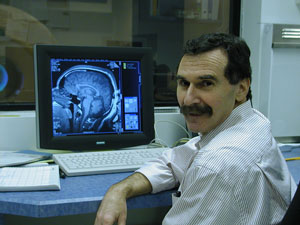 Dr. Elliot Stein, chief of the Neuroimaging Research Branch, within NIDA's Intramural Research Program, says functional magnetic resonance imaging makes it possible to see the impact of drugs on the brain at work.
"Using functional magnetic resonance imaging [fMRI], IRP researchers will be able to make observations that are not possible in any other type of study," he says. "The technique doesn't require use of radioactive compounds as PET imaging does, and it doesn't use radiation to create an image as conventional x-ray or CT does.
"We can use fMRI to actually watch the brain at work and see what regions are active while participants perform cognitive tasks or are presented images or other environmental cues related to drug abuse," continues Dr. Stein. "This provides quantitative measurements of functional activity for different areas of the brain--not just paper-and-pencil scores or ratings on a 1-to-10 scale--to help us understand a drug's effect on reasoning, anticipation, or reward. Does the brain work differently, in some fundamental way, when a person is using drugs? When she or he is abstinent or going through withdrawal?"
Moreover, fMRI studies are not limited to investigating the effects of drugs. They can also be used to evaluate treatment, notes Dr. Stein. "If we understand the effect of a drug on how a patient learns, remembers, or pays attention, we can try to develop behavioral treatments that focus on those functions.
"By watching the brain perform the same tasks before and after treatment, we literally can see if there is a functional effect of the treatment and use that information to determine if it is more likely than other treatments to help the patient. Using fMRI to guide development of treatments and evaluate their impact will make it possible to tailor a behavioral treatment to how a patient's brain actually works."
Heads Up Poster Winner Selected

NIDA and Scholastic Classroom Magazines unveiled the Grand Prize artwork from the national Heads Up: Real News About Drugs and Your Body poster contest on May 16 at Scholastic Headquarters in New York City. The contest was part of the ongoing Heads Up science-based drug education campaign, for which NIDA and Scholastic partnered.
Eighth-grader Ania Lisa Etienne, from Brooklyn, New York, created the winning poster. Selected from nearly 1,100 entries, Ania's poster features a distressed teenager looking into a shattered mirror, a symbol of the bad luck that results from drug abuse, Ania explains. Her winning artwork and slogan, "You Can't Sniff Away Your Sorrows," will form the basis of a poster to be included in the Heads Up program during the 2003-2004 school year.