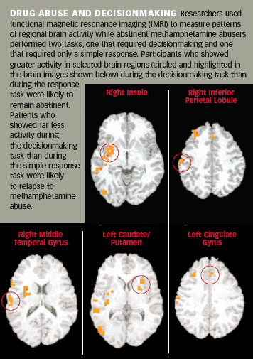
NIDA-supported investigators have found that functional magnetic resonance imaging (fMRI) of the brain, performed during a psychological test, can predict with high accuracy whether an individual will relapse following treatment for methamphetamine abuse. Their study revealed a characteristic pattern of brain activity in methamphetamine-abusing men who relapsed within 1 to 3 years after completing treatment and a different pattern in men who did not.

Dr. Martin Paulus and colleagues at the University of California, San Diego, took the point of departure for their work from previous research that showed methamphetamine abusers and nonabusers activating different brain areas during psychological tests of decisionmaking. These earlier studies showed that poor choices made by drug abusers correlate to distinctive patterns of activity in some areas of the brain. Dr. Paulus's team hypothesized that activity patterns in those regions might also be associated with relapse to drug abuse, which involves similarly destructive decisions.
To test their hypothesis, the researchers recruited 46 men who had voluntarily entered and completed a 28-day inpatient drug treatment program after abusing methamphetamine for periods ranging from 3 to 34 years. When each man had been abstinent for about 4 weeks, he participated in two psychological tests. During one, he was asked to watch a computer screen and press a button every time a symbol appeared. In the other, he was asked to try to predict whether a flashing symbol would next occur on the left side or right side of the computer screen. The difference between the two tasks was that, in the first, the test-taker needed only to react upon seeing the symbol, while in the second, he needed to decide which side to choose. The researchers recorded the men's brain activity with fMRI throughout the tests.
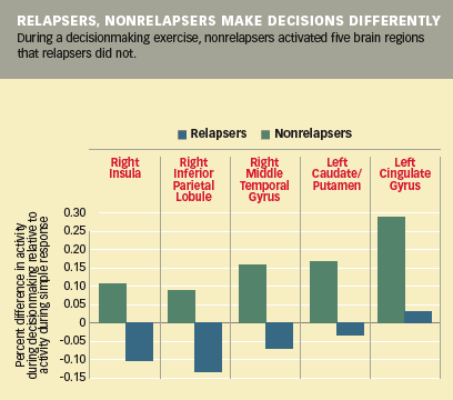
A year or more (360 to 967 days) after the imaging sessions, Dr. Paulus's team was able to locate and contact 40 of the 46 patients. Of these, 18 had relapsed to methamphetamine abuse (median time to relapse, 279 days; range, 36 to 820 days). Comparing their fMRI results with those of the 22 nonrelapsers, the researchers noted nine regions where the groups' brain activity had differed during decisionmaking. The relapse group showed less activation of the dorsolateral, prefrontal, parietal, and temporal cortices and the insula—regions associated with evaluation and choice among actions that may lead to either beneficial or harmful outcomes. The patterns of brain activation predicted relapse in 17 of the 18 men who had resumed methamphetamine abuse and predicted successful abstinence in 20 of the 22 patients who had not relapsed, Dr. Paulus says.
"The most striking aspect of this result is that the fMRI pattern has 90 percent accuracy in predicting outcome," Dr. Paulus says. "The differences in brain activity are pronounced, with little overlap." Differences in the right insula, right posterior cingulate, and right middle temporal gyrus differentiated relapsers from nonrelapsers. Other brain regions predicted the timing of relapse.
"Some of these predictive areas have not previously been strongly associated with drug abuse," observes Dr. Steven Grant of NIDA's Division of Clinical Neurosciences and Behavioral Research. "For example, while other investigators have reported alterations in the parietal lobe related to drug abuse, this is the first study to show the parietal cortex playing an important role. However, because so many brain regions were related to relapse, we still do not have a full understanding of what specific process might be dysfunctional in the relapse group."

The potential clinical implications of the new finding are promising, but uncertain. For example, no women were included among the participants, who were enrolled from treatment programs. "It's important to confirm the findings in women, for whom social, demographic, and other factors associated with relapse may differ," Dr. Paulus points out. Nonetheless, he says that, in principle, programs treating methamphetamine abuse might use the fMRI protocol to assess patients, then assign those likelier to relapse to higher levels of care. Dr. Paulus believes such an approach might prove cost-effective, even with typical fMRI charges of up to $700 per hour in academic imaging centers. "The human and social costs of relapse are high," Dr. Paulus says. "Using this imaging technique to precisely allocate care to the patients who need it most might well produce enough savings elsewhere to more than offset its expense. An alternative, more practical course of action might be to use these fMRI results as a benchmark for development of other assessments that are less costly, but have the same predictive strength."
Source
- Paulus, M.P.; Tapert, S.F.; and Schuckit, M.A. Neural activation patterns of methamphetamine-dependent subjects during decision making predict relapse. Archives of General Psychiatry 62(7):761-768, 2005. [Abstract]
