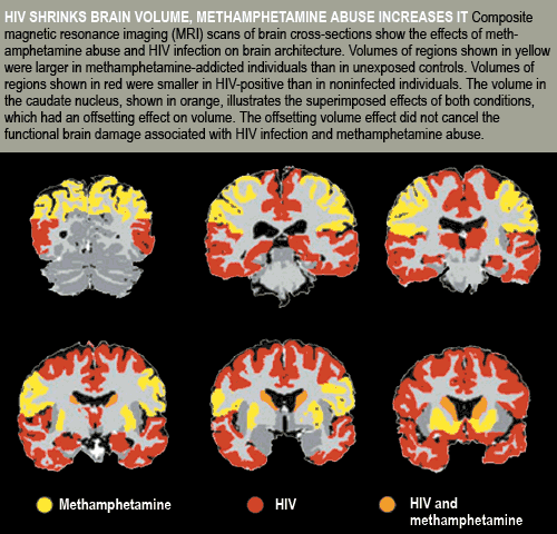
In a study that confirmed the association between HIV infection and loss of brain volume, NIDA-funded investigators also found an association between methamphetamine addiction and increased regional brain volume. Each type of volume change was associated with neurocognitive impairments, but it was unclear whether the two together caused any cognitive effects beyond the sum of what each produced individually.
Using structural magnetic resonance imaging (MRI), Drs. Terry L. Jernigan, Anthony C. Gamst, and colleagues at the University of California, San Diego (UCSD) mapped the major gray-matter brain structures of 103 people in four ageand education-matched groups: HIV-infected; methamphetamine-addicted; having both conditions; and having neither. The methamphetamine-addicted individuals were in recovery at UCSD's HIV Neurobehavioral Research Center.
After accounting for normal age-related reductions in brain volume, the participants with HIV had smaller volumes of cortical, limbic, and striatal structures, with the associations being most pronounced in the frontal and temporal lobes. Methamphetamine addiction was linked with increased volume in the parietal cortex and in all three segments of the basal ganglia—caudate nucleus, lenticular nucleus, and nucleus accumbens (NAc). In the caudate, volume reductions related to HIV and increases related to methamphetamine overlapped, producing a net volume approximating normal.

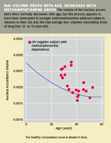
A further analysis of the volume data may reinforce existing evidence that drug abuse is especially damaging during adolescence and young adulthood, when the brain is still developing. The results showed that addicted individuals who were younger had greater NAc volume differentials compared with their non-drug-abusing age mates than did older addicted individuals. One possible explanation for this is that the drug interfered with the pruning of some NAc connecting fibers that normally occurs in the transition to adulthood, producing a small but measurable reduction in NAc volume. "While we can't be certain of the explanation, this finding highlights the concern that exposure during adolescence may alter the course of ongoing brain maturation," says Dr. Jernigan (see chart below).

Although the study results provided little information about specific drug mechanisms, the investigators note that animal studies have shown methamphetamine can incite inflammatory responses and abnormal growth of nerve fibers, each of which can increase tissue volume. "These findings emphasize that the brain's response to stimulant exposure, and indeed to HIV as well, is probably quite dynamic, characterized by overlapping responses in different glial, as well as neuronal, cell populations," says Dr. Jernigan. "The findings raise interesting questions for multiple-modality imaging studies, and underscore the degree of neural plasticity, and thus the potential for targeted intervention." Dr. Jernigan says a finding of extensive change in the parietal cortex of methamphetamine abusers "helps to confirm the importance of parietal lobe involvement and may help correct a tendency in the field to neglect this region."
Implications for Brain Function
The researchers looked for correlations between the brain volume abnormalities and ratings of neuropsychological impairment. At the outset of the study, all other groups were significantly impaired relative to the HIV-negative and methamphetamine-negative group, which had a rating of 2.9 compared with 4.7 for those with both conditions, 4.2 for methamphetamine-addicted participants and 4.1 for HIV-positive individuals. Brain impairment was most pronounced in the HIV-positive participants with the most extensive loss of cortical volume and in the methamphetamine-addicted participants with the highest increase in cortical volume. The investigators found only one significant correlation between brain volumes and impairment in addicted individuals with HIV, a finding they believe probably reflects confounding by the opposed volume impacts of the two pathologies. The correlation was between hippocampal volume—a structure that both factors may damage—and severity of cognitive impairment in the dually diagnosed group.
"The fact that brain alterations in methamphetamine dependence and HIV infection are distinct from each other is a clue that may help us to sort out the origins of different kinds of mental problems in these individuals," Dr. Jernigan says." This is very exciting, because our results raise a number of specific questions that may not have been posed without these findings."
Dr. Ro Nemeth-Coslett, a NIDA psychologist, agrees. "As often happens in research, these results raise more questions than they answer. Dr. Jernigan's findings of structural inconsistencies in pathology are unaccounted for. Now we need mechanistic studies to provide a clearer understanding of what aspects of microscopic cellular organization actually drive the MRI measures."
Source
- Jernigan, T. L., et al. Effects of methamphetamine dependence and HIV infection on cerebral morphology. American Journal of Psychiatry 162(8):1461-1472, 2005. [Abstract]
