People who have used cocaine for a long time report a paradoxical-seeming experience: The pleasure they get from taking the drug decreases, even as the drug intensifies its hold over their behavior.
A recent NIDA-supported study sheds light on why this might be. Researchers showed that, in mice, a cocaine-induced imbalance in the activity of two key populations of neurons in the reward system persists for a longer period after repeated exposure to the drug. For long-term users, the researchers suggest, this change could both weaken the cocaine “high” and strengthen the compulsion to seek the drug.
A Distorted Ratio
Drs. Congwu Du and Yingtian Pan and colleagues at Stony Brook University in New York and at NIDA injected two groups of mice with a single dose of cocaine (8 milligrams per kilogram of body weight). One group of mice had already been exposed to the drug daily for 2 weeks, and the other, a control group, was receiving the drug for the first time. Using a novel dual-imaging and measurement technique (see Lights, Camera, Neuron Action), the researchers tracked the drug’s impact on activity levels of two populations of medium spiny neurons (MSNs) in the striatum of the two groups.
One of two MSN populations observed by the researchers interacts with dopamine via receptors called D1R. When activated by dopamine, striatal D1R MSNs give rise to pleasurable feelings, motivate an animal or a person to repeat the experience that yielded these feelings, and promote the conversion of such motivation into action by stimulating neurons in the brain’s motor cortex. The other MSN population interacts with dopamine via a different receptor, called D2R. When activated, D2R MSNs counter the effects of the D1R MSNs. They attenuate euphoria and drug seeking and inhibit the motor cortex.
In the experiment, Drs. Du and Pan found that immediately after the cocaine injection, D1R MSN activation increased and D2R MSN activation decreased, both in the mice that had been exposed daily to the drug and in those being exposed for the first time. As a result, in both groups, the ratio of D1R MSN to D2R MSN activation shifted sharply in favor of the D1R MSNs and their reward- and motivation-promoting effects.
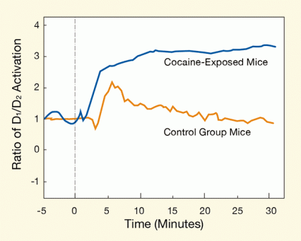
At 5 to 7 minutes post-injection, however, the D1R/D2R activity ratios diverged between the two groups of mice. In the control mice, D1R activation rapidly fell back to its baseline level, causing the D1R/D2R ratio to also return to near baseline. In the mice that had been exposed daily to cocaine, in contrast, the cocaine-induced D1R activation increased steadily over the entire 30 minutes that the animals were observed. As a result, D1R/D2R rose higher and remained elevated longer in the daily-exposed mice than in the control mice (see Figure 1).
Drs. Du and Pan note that the rapid rise and fall of the D1R/D2R MSN activity ratio seen in control mice conforms to a “fast-in, fast-out” pattern that has been shown to underlie the exaggerated euphoria that cocaine and other addictive drugs confer. The researchers suggest that if the departure from this activation pattern that they observed in the daily-exposed mice holds in people as well, it could help explain why long-term users of cocaine report less euphoria from taking the drug.
The researchers propose that the drawn-out time course of cocaine-induced D1R/D2R MSN activation following repeated exposure will also enhance an animal’s or a person’s drive to seek the drug. Dr. Pan explains, “Dopamine both activates and inhibits brain circuits, and normally this dual action produces healthy behavioral outcomes. Cocaine upsets this balance. It enhances the D1R MSN signaling that promotes drug-seeking behaviors in people and animals, and suppresses D2R MSN signaling that normally puts a brake on those behaviors. In our experiment, we showed that this imbalance is short lived in mice when they are exposed to the drug for the first time, but long lasting in mice that have already been repeatedly exposed.”
 Figure 1. Regular Cocaine Exposure Skews the D1R/ D2R Signaling Toward Reward Signaling In both cocaine-naïve and daily-exposed mice, the D1R/ D2R ratio increased abruptly after a cocaine injection. In the cocaine-naïve mice, the ratio quickly returned to near-baseline, but in the daily-exposed mice, it remained elevated over 30 minutes of observation.
- Text Description of Figure 1
-
The figure shows a line graph of the ratio of the D1 and D2 receptor activations. The vertical (y) axis shows the ratio of D1/D2 activation in units ranging from −1 to 4, and the horizontal (x) axis shows the time in minutes of the experiment; the axis starts at −5 minutes before the cocaine injection (marked by a vertical dashed line at 0 minutes) to 30 minutes. As shown by a blue line, about 2 minutes after the cocaine injection, in the cocaine-exposed mice, the D1/D2 activation ratio quickly rises from a baseline level of 1 at 0 minutes to about 2.5 at 4 minutes and reaching a peak activation ratio of about 3 after about 10 minutes. The line than stays at that level for the remaining 20 minutes recorded. As shown by an orange line, about 3 to 4 minutes after the cocaine injection, in the cocaine-naïve mice, the D1/D2 activation ratio rises from a baseline level of 1 to a peak activation ratio of about 2 at about 7 minutes, after which the ratio drops steadily back to baseline in the remaining 23 minutes recorded.
More Research Is Needed
“The research field had not put much effort into separating how these two dopamine receptor systems are involved in rewiring the brain exposed to chronic cocaine use or their effects on compulsive intake of drug,” says Dr. Nora D. Volkow, NIDA Director and a collaborator on the study. “This work highlights the importance of the relative participation of D1R versus D2R signaling.”
Drs. Du and Pan have more work to do to show that their observations account for long-term cocaine users’ reduced enjoyment and increased compulsion to use the drug. As a first step, they plan to examine whether increasing the D1R/D2R MSN activity ratio indeed increases animals’ drug-seeking behavior. This will be very challenging, says Dr. Du, because it will require adapting their imaging technique to monitor MSNs in awake and moving animals. To date, they have used it only with anesthetized and restrained animals.
Another outstanding question is whether long-term cocaine use actually changes the time course of D1R MSN activation in people as it does in mice. Dr. Pan notes that although research has not yet addressed this question, imaging studies conducted in Dr. Volkow’s laboratory have shown that cocaine dampens D2R signaling in people as well as in mice.
If further investigations confirm the researchers’ hypotheses, says Dr. Volkow, “treatments that strengthen D2R signaling could help people stop using cocaine.”
This study was supported by NIH/NIDA grants DA021200, DA028534, DA032228, and DA029718, with collaboration from Dr. Nora Volkow in her role as an intramural scientist at the National Institute on Alcohol Abuse and Alcoholism.
Source:
Park, K.; Volkow, N.D.; Pan, Y.; Du, C. Chronic cocaine dampens dopamine signaling during cocaine intoxication and unbalances D1 over D2 receptor signaling. J. Neurosci. 33(40):15827-15836, 2013. Full Text
Lights, Camera, Neuron Action
To achieve their observations, Drs. Du and Pan developed a dual-imaging technique based on a novel microprobe that was used for visualizing individual neurons deep within the brain. The technique enabled them to distinguish the populations of D1R and D2R MSNs, and to track moment-to-moment changes in each one’s calcium levels. Calcium levels directly reflect a neuron’s level of activation.
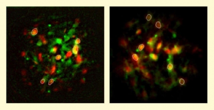
Dr. Du recounts, “First, we used genetically engineered mice to incorporate visual markers called enhanced green fluorescent proteins (EGFPs) in either their D1Rs or their D2Rs. When we exposed an animal’s striatum to light from a fluorescence microscope, the EGFPs would emit green light, revealing whichever population had been tagged. Next, we infused each animal’s striatum with a calcium indicator, called Rhod2, which emits red light when it binds to calcium after excitation under a fluorescent microscope and increases in brightness when the calcium level is increased. Then, we performed continuous imaging through the fluorescence microscope before, during, and for 30 minutes after administering cocaine. Finally, we combined and analyzed the images in a computer.” (See Figure 2.)
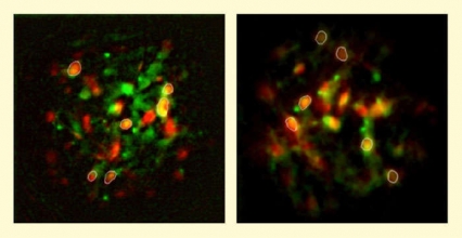
 Figure 2. Images Used to Measure and Compare Activation of D1R (Left) and D2R (Right) MSNs in the Striatum The researchers used mice in which either D1R or D2R MSNs had been labeled with EGFP and used Rhod2 dye to visualize calcium levels in the cells. With a technique called microprobe fluorescent imaging, the researchers took images of fluorescent EGFP cells and of the fluorescent Rhod2 dye in the mice’s striatum after the animals had received cocaine. They then combined the two images to select EGFP cells (showing a green glow) and to measure levels of Rhod2 (showing a red glow and serving as proxy for calcium levels) in these cells; cells selected for the measurements are circled.
- Text Description of Figure 2
-
The figure shows two fluorescent microscopy images of medium spiny neurons that express enhanced green fluorescent protein (resulting a green glow in cells), indicating expression of either the D1 or the D2 receptor on the MSNs, and show staining with rhodamine dye, which shows a red glow when bound to calcium. The left image shows neurons expressing the D1 receptor, and the right image shows neurons expressing D2 receptors. MSNs showing both green and red glows are circled in white and both expressed EGFP, indicating D1R or D2R expression, and showed rhodamine fluorescence, used to determine calcium levels for measuring MSN activation.

