
This research:
- Demonstrated that use of anabolic-androgenic steroids (AAS) is associated with compromised heart pumping and atherosclerotic plaque.
- Suggests that clinicians should inquire about AAS use when young or middle-aged men present with left ventricular dysfunction or coronary artery disease.
An estimated 3 million to 4 million Americans have used anabolic-androgenic steroids (testosterone or synthetic derivatives of testosterone) to gain muscle mass for sports or to enhance their appearance. A large new study demonstrates that such use can narrow the coronary arteries and impair left ventricular (LV) function. Together, these effects can reduce the supply of oxygenated blood both to the heart and from the heart to the arteries.
Dr. Aaron Baggish and colleagues at Massachusetts General Hospital in Boston and McLean Hospital in Belmont, Massachusetts, assessed the heart and coronary arteries in 140 male weightlifters ages 34 to 54. All the men could bench-press 275 pounds at some point in their lives; 58 were currently using AAS; 28 had previously used AAS for at least 2 years; and 54 had never used AAS. Compared with the nonusers, the AAS users had higher mean blood pressure and prevalence of elevated low-density lipoprotein (“bad”) cholesterol, which is a risk factor for atherosclerosis.
Echocardiography revealed that 71 percent of current AAS users had a lower-than-normal LV ejection fraction, a measure of how much oxygenated blood the heart delivers to the arteries with each contraction (see Figure 1). Half of current AAS users exhibited slower-than-normal LV expansion, indicating that reduced amounts of oxygenated blood flowed in to refill the LV during the relaxation phase of the heartbeat. These impairments likely were related to thickening of the LV muscle wall in the AAS users. Former AAS users had a larger LV ejection fraction than current users, suggesting that their LV had recovered some pumping strength.
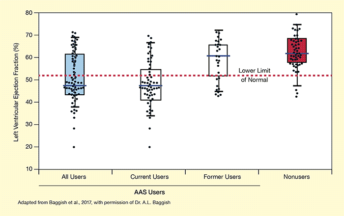 Figure 1. Long-Term Anabolic-Androgenic Steroid (AAS) Use Damages Heart Function Among a group of weightlifters, AAS users’ left ventricular ejection fractions (LVEF) were lower than those of nonusers and often below the normal range, suggesting impaired heart pumping capacity. Current AAS users had lower LVEF than former users, suggesting that some recovery may occur after quitting.
In this boxplot, black dots represent individual weightlifters’ LVEF. All LVEF values below the horizontal red line are below the normal range, suggesting compromised heart pumping capacity. The horizontal line inside the box for each group represents the median LVEF in the group. The top and bottom lines of the boxes represent the 75th and 25th percentile LVEF in each group. Dots outside of the horizontal lines above and below each box represent weightlifters with extremely high or low LVEF, based on the overall range of LVEF in that group.
- Text Description of Figure 1
-
The figure shows a box diagram of the left ventricular ejection fraction (LVEF) of weightlifters who were users and nonusers of anabolic-androgenic steroids (AAS). The vertical (y-)axis shows the LVEF in percent. A horizontal red dotted line at about 53% indicates the lower limit of normal for LVEF. The horizontal (x-) axis shows the four groups of participants analyzed, including, from left to right, all AAS users, current AAS users, former AAS users, and nonusers who had never used AAS. Dots in each group indicate the measured values for individual participants. For each group, the box indicates the range from the 25th percentile (bottom edge) to the 75th percentile (top edge) of all measured values within that group, with the horizontal line in the box indicating the median. Vertical bars for each group indicate the range of measures in that group. For each group, the lower end of the vertical bar indicates the minimum calculated LVEF and the upper end of the vertical bar indicates the maximum calculated LVEF. Dots beneath the lower end of the bar or above the upper end of the bar are considered outliers (i.e., extremely low or high LVEF for that group). For all AAS users (left group), the lower limit of the LVEF is about 36%, the 25th percentile is about 43%, the median is about 48%, the 75th percentile is about 61%, and the maximum is about 70%. For current AAS users, the minimum LVEF is about 34%, the 25th percentile is about 41%, the median is about 48%, the 75th percentile is about 55%, and the maximum is about 68%. For former AAS users, the minimum LVEF is about 45%, the 25th percentile is about 53%, the median is about 60%, the 75th percentile is about 65%, and the maximum is about 72%. For nonusers, the minimum LVEF is about 46%, the 25th percentile is about 57%, the median is about 62%, the 75th percentile is about 69%, and the maximum is about 75%.
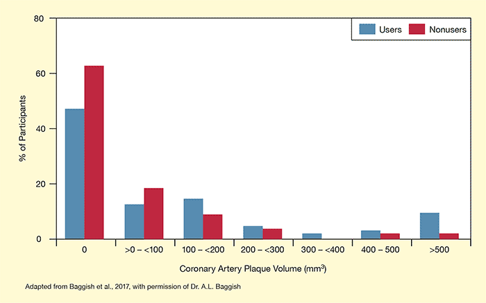 Figure 2. AAS Use Increases Atherosclerotic Plaque in the Coronary Arteries AAS users were less likely to have no deposits of atherosclerotic plaque and more likely to have larger plaque deposits in their coronary arteries than did nonusers. Plaque obstructs blood flow to the heart and large-volume deposits can increase risk of heart disease.
- Text Description of Figure 2
-
The bar chart shows the coronary artery plaque volumes in AAS users and nonusers. The horizontal (x-) axis shows different categories of plaque volume in mm3, including, from left to right, 0 mm3, more than 0 mm3 to less than 100 mm3, 100 mm3 to less than 200 mm3, 200 mm3 to less than 300 mm3, 300 mm3 to less than 400 mm3, 400 mm3 to 500 mm3, and more than 500 mm3. The vertical (y-) axis shows the percentage of participants in each category, from 0% to 80%. For each category, AAS users are represented by blue bars and nonusers are represented by red bars. About 50% of users and 62% of nonusers had no coronary artery plaque. More than 0 mm3 but less than 100 mm3 of plaque was found in about 15% of AAS users and 20% of nonusers. From 100 mm3 to less than 200 mm3 of plaque was found in about 18% of AAS users and 10% of nonusers. From 200 mm3 to less than 300 mm3 of plaque was found in approximately 5% of AAS users and 4% of nonusers. From 300 mm3 to less than 400 mm3 of plaque was found in about 2% of AAS users and 0% of nonusers. From 400 mm3 to 500 mm3 of plaque was found in about 3% of AAS users and 2% of nonusers. More than 500 mm3 of plaque was found in about 11% of AAS users and 2% of nonusers.
AAS users also had significantly more atherosclerotic plaque in their coronary arteries than did nonusers (see Figure 2). This effect was more pronounced the longer they had taken AAS and did not seem to be reversible after discontinuing AAS use. Coronary atherosclerotic plaque obstructs blood flow to the heart muscle, increasing the risk of ischemia, heart attack, and heart failure. Notably, three AAS users, but none of the nonusers, had suffered heart attacks due to coronary artery disease at relatively young ages (38, 43, and 46 years). Another user experienced heart failure with underlying coronary artery disease at age 42.
The researchers found no differences in cardiovascular structure and function between AAS nonuser weightlifters and nonusers who were not weightlifters. This observation confirmed that none of the observed changes could be attributed to weightlifting itself.
Dr. Harrison G. Pope, one of the study’s primary researchers, concludes, “This study provides strong evidence that anabolic steroids pose serious and sometimes even fatal dangers to the muscle and the blood vessels of the heart. Given that some 3 million Americans have tried these drugs, this represents a significant public health problem.”
Dr. Jag H. Khalsa, Chief of NIDA’s Medical Consequences of Drug Abuse and Co-Occurring Infections Branch, agrees, pointing out that the findings contradict the general perception that AAS use is safe. “It is of paramount importance that clinicians are made aware of this highly significant finding so that they can advise their patients about the dangers of anabolic steroid use,” he emphasizes.
This study was supported by NIH grant DA029141.
Source:
Baggish A.L., Weiner, R.B., Kanayama, et al. Cardiovascular toxicity of illicit anabolic-androgenic steroid use. Circulation 135(21):1991-2002, 2017.