A new study adds to the copious existing evidence that chronic exposure to addictive drugs alters the brain in ways that make quitting difficult. NIDA-supported researchers showed that, in monkeys, methamphetamine alters brain structures involved in decision-making and impairs the ability to suppress habitual behaviors that have become useless or counterproductive. The two effects were correlated, indicating that the structural change underlies the decline in mental flexibility.
Human chronic methamphetamine users have been shown to differ from nonusers in the same ways that the post-exposure monkeys differed from their pre-exposure selves. The researchers’ use of monkeys as study subjects enabled them to address a question that human studies cannot: Did the drug cause those differences, or were they present before the individuals initiated use of the drug? The study results strongly suggest that the drug is significantly, if not wholly, responsible.
Before and After Methamphetamine
A defining feature of addiction to methamphetamine is that the addicted individual keeps on taking the drug despite the negative health and social effects of doing so. Psychological testing of addicted individuals has linked their difficulty quitting to a weakness in inhibitory control—a reduced ability to stop repeating previously learned behaviors. Brain imaging studies have also shown that, compared to nonusers, chronic users of the drug have, on average, more gray matter in the putamen and less gray matter in the prefrontal cortex (PFC) brain regions.
To clarify the relationships among these observations, Dr. David Jentsch, graduate students Stephanie Groman and Angelica Morales, and colleagues at the University of California, Los Angeles (UCLA) exposed 7 adult male vervet monkeys to methamphetamine in a 31-day regimen of escalating doses that simulates chronic use of the drug in humans. The researchers tested the monkeys’ cognition before, during, and after the methamphetamine exposure, and obtained magnetic resonance images (MRI) of the brain before and after the exposure.
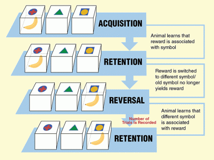
Before methamphetamine exposure, the experimental monkeys performed as well on a test of inhibitory control as 7 control monkeys that received only saline injections. When retested after 3 weeks on the methamphetamine regimen, their performance had declined significantly. In the test, the researchers trained the animals to point to one of three symbols to receive a fruit reward, then switched the reward to another symbol. The methamphetamine-exposed monkeys took many more tries to shift from pointing to the first symbol to pointing to the other (see Figure 1).
Similarly, the MRIs of the experimental monkeys’ putamen matched those of the control monkeys’ prior to methamphetamine exposure, but differed after exposure. Analysis of the images revealed significant increases in gray matter in the right putamen.
The researchers hypothesized that the expansion of putamen gray matter could explain the monkeys’ compromised test performance. A primary role of the putamen is to initiate established or habitual responses to familiar situations or stimuli. In the normal shaping of behavior, other brain structures, in particular the PFC, functionally inhibit the putamen from initiating those responses in circumstances where they are inappropriate. However, an enlarged putamen may override this input and trigger habitual responses even when they are useless or harmful.
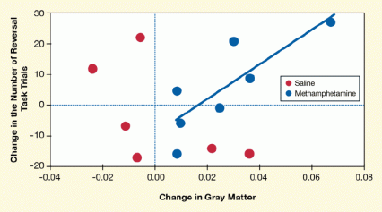
To test this hypothesis, Dr. Jentsch and colleagues plotted the experimental monkeys’ decline in performance on the test of inhibitory control against their increase in putamen gray matter. The results were confirmatory: The animals that showed the greatest increases in putamen size were slowest to adjust to the altered reward structure (see Figure 2).
“When the brain’s structural integrity is dysregulated in the striatum [the brain region that contains the putamen], we think the animal’s behavior is unleashed from inhibitory control,” Dr. Jentsch says.
Dr. Jentsch notes that reduced gray matter in the PFC, as occurs in human methamphetamine users, would be expected to further weaken inhibitory control over habitual responses. Although the researchers anticipated that methamphetamine would cause contraction of the experimental monkeys’ PFC, there was no statistical change measured in the study, possibly because of the relatively small number of monkeys involved in the study. Alternatively, he adds, there is some evidence that changes in PFC observed in humans result from other habits associated with methamphetamine use, such as tobacco smoking.
Can the Brain Recover?
The UCLA study’s findings underscore the importance of the PFC–putamen circuitry in methamphetamine addiction. “These results provide a causal mechanism for why methamphetamine addiction is so hard to treat and has a significant chance of relapse early in treatment,” says Dr. James Bjork of NIDA’s Clinical Neuroscience Branch. “It’s a double whammy if the part of the brain that normally controls risky behavior is weakened while the habit-sustaining center is chemically beefed up and on overdrive.”
With their impact on behavior, these brain centers constitute potential prime targets for interventions to help people regain control over their drug use. Such interventions could range from medications that correct abnormalities in neurotransmitters and other chemicals in these brain regions to aerobic exercise and training of self-control, both of which may alter PFC–putamen structure and function.
“Going forward,” Dr. Jentsch says, “questions that need to be answered are: What are the cellular and molecular events in the striatum that initiate the whole chain of structural and behavioral changes?” Last year, the UCLA researchers turned up a possible clue: In a study using positron emission tomography (PET) imaging, methamphetamine-exposed monkeys had fewer dopamine transporters and D2 receptors in the putamen and related brain structures, compared to unexposed monkeys.
Another topic on Dr. Jentsch’s research agenda is the source of individual differences in drug use. Among the drug-exposed monkeys in their trial, for example, increases in putamen volume ranged from less than 2 percent in one animal to 11 percent in another, with corresponding differences in performance on the test of inhibitory control. This mirrors the circumstance among human chronic users of the drug.
"Where are these individual differences in drug use coming from? What makes the drug-susceptible brains different from the resilient brains?” Dr. Jentsch asks. “These questions are important because the answers will enable us to identify individuals with greater risk for being damaged by methamphetamine, and target them with preventive efforts that include educating them about the potential impact of the drug on their brains.” As a first step in this quest, the UCLA team has fully sequenced the genomes of the colony of vervet monkeys from which these study subjects were obtained. They will analyze these data for gene variants that associate with the observed differences in drug responses.
Ultimately, can methamphetamine’s disruptive effects on the brain be reversed? In the UCLA study, the methamphetamine-induced changes persisted through the final cognitive and behavioral assessments, which occurred 3 weeks after the animals received their last dose of the drug. Therefore, Dr. Jentsch says, to answer this question, future studies should track primate brains through longer abstinence.
“Methamphetamine dependence is currently a problem with no good medical treatments,” Dr. Jentsch notes. “When you say a disease like methamphetamine dependence is costly, it’s not just costing money, but lives, productivity, happiness, and joy. Its impact bleeds through families and society.”
This study was supported by NIH grants DA022539, DA020598, DA024635, DA028812, and DA033117.
- Text description of Figure 1
-
The graph shows a flow chart depicting the four stages of a reversal trial, which measures an animal’s mental flexibility. A piece of fruit (a banana) is placed as a reward into one of three boxes labeled with different symbols on their lids (a red cross, green triangle, or yellow sphere). During the acquisition and retention phases (shown on first and second lines from the top) an animal learns to retrieve the fruit reward from one of the boxes marked with a specific symbol (i.e., the red cross). In the reversal and second retention phases (third and fourth lines from the top), the animal has to learn that the fruit reward is no longer available from the box with the cross symbol, but in a box with a different symbol (i.e., the yellow sphere). The number of trials it takes the animal to successfully associate the fruit reward with the new symbol during the second retention phase is recorded to measure the animal’s ability to adapt to a new reward cue.
- Text description of Figure 2
-
The figure shows a scatter plot indicating the relationship between the number of reversal trials, i.e., the number of attempts a vervet monkey made to obtain a food reward after it had been switched to a new visual cue, and a change in gray matter in the monkeys’ brains after exposure to methamphetamine or to saline (an inactive control). The vertical (y)-axis shows the change in the number of reversal trials, which was calculated by subtracting the number of trials before each exposure from the number of trials after the exposure. A positive number for the change indicates that an animal needed a greater number of trials to obtain the food reward after being exposed to methamphetamine or saline and a negative number that it needed fewer trials. The horizontal (x)-axis shows the change in gray matter, which was measured by comparing signal intensities in magnetic resonance imaging scans before and after the monkeys were exposed to the drug or saline. As shown in the graph, after methamphetamine exposure (blue circles), most monkeys needed more trials to obtain the food reward than the monkeys exposed to saline (red circles); most saline-exposed monkeys required fewer trials than before the exposure. The worsening performance of the methamphetamine-exposed monkeys was correlated (indicated by the solid blue line) with increased gray matter in their right putamen.
Sources
Groman, S.M.; Morales A.M.; Lee, B.; London, E.D.; Jentsch, J.D. Methamphetamine-induced increases in putamen gray matter associate with inhibitory control. Psychopharmacology 229(3):527-538, 2013. Abstract
Groman, S.M.; Lee, B.; Seu, E.; James, A.S.; Feiler, K.; Mandelkern, M.A.; London, E.D.; Jentsch, J.D. Dysregulation of D2-mediated dopamine transmission in monkeys after chronic escalating methamphetamine exposure. The Journal of Neuroscience 32(17):5843–5852, 2012. Full text


