
The objective of the presentation is to inform students (high school) how 3 drugs of abuse (cocaine, opiates, marijuana) actually work in the brain. The presentation is arranged in 3 sections. The first section introduces the brain and presents some basic neurobiology, the second introduces the reward pathway and the third presents the mechanism of action of each drug and how each affects the reward system.
This presentation can be downloaded as a Powerpoint file - The Brain & the Actions of Cocaine, Opioids, and Marijuana (PPT, 4.1MB) and was last reviewed in November, 2019
Introduction to the Brain
- Introduction
-
Image
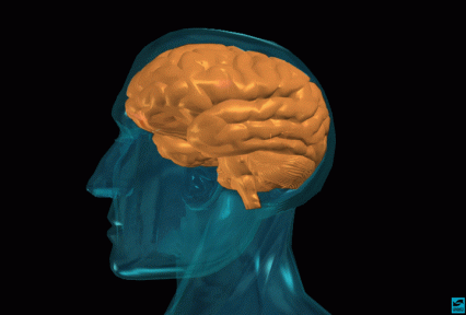
Introduce the topic of your talk. Indicate that you will explain how the brain basically works and how drugs such as cocaine, opioids and marijuana interact with the brain's normal activities. Tell students that you will introduce the concept of "reward" which is the property that is characteristic of many addictive drugs. Describe the brain as a functional unit; it is made up of billions of nerve cells (neurons) that communicate with each other using electrical and chemical signals.
- Brain regions and neuronal pathways
-
Image
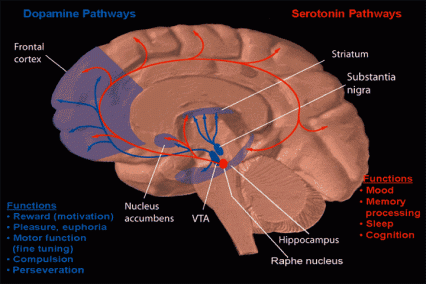
Certain parts of the brain govern specific functions. Point to sensory, motor, association and visual cortex to highlight specific functions. Point to the hippocampus to highlight the region that is critical for memory, for example. Indicate that nerve cells or neurons travel from one area to another via pathways to send and integrate information. Show, for example, the reward pathway. Start at the ventral tegmental area (VTA) (in blue), follow the neuronal path to the nucleus accumbens (purple), and then on to the frontal cortex. Explain that this pathway gets activated when a person receives positive reinforcement for certain behaviors ("reward"). Indicate that you will explain how this happens when a person takes an addictive drug.
- Neuronal structure
-
Image
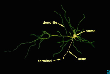
Remind the student that pathways are made up of neurons. Describe the anatomy of a neuron (soma, dendrites, and axon are marked with text). State that this neuron is real - as viewed through a microscope. Explain the normal direction of impulse flow. Dendrites and soma receive chemical information from neighboring neuronal axons. The chemical information is converted to electrical currents which travel toward and converge on the soma. A major impulse is produced (the action potential) and travels down the axon toward the terminal. Point to the terminal.
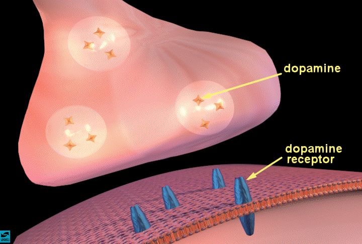
- The synapse and synaptic neurotransmission
-
Image
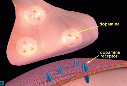
Describe the synapse and the process of chemical neurotransmission. Indicate how vesicles containing a neurotransmitter, such as dopamine (the stars), move toward the presynaptic membrane as an electrical impulse arrives at the terminal. Describe the process of dopamine release (show how the vesicles fuse with the presynaptic membrane). Once inside the synaptic cleft, the dopamine can bind to specific proteins called dopamine receptors (in blue) on the membrane of a neighboring neuron. Introduce the idea that occupation of receptors by neurotransmitters causes various actions in the cell; activation or inhibition of enzymes, entry or exit of certain ions. State that you will describe how this happens in a few moments.
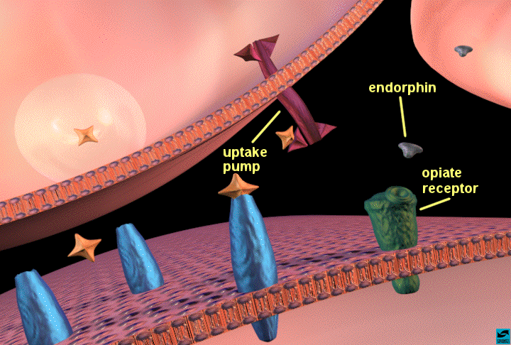
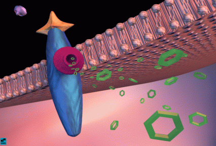
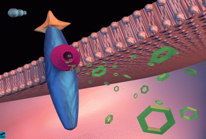
- Dopamine neurotransmission
-
Image
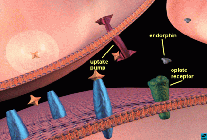
Using the close-up of a synapse, continue using dopamine for your example of synaptic function. Explain that it is synthesized in the nerve terminal and packaged in vesicles. Reiterate the steps in neurotransmission. Show how the vesicle fuses with the membrane and releases dopamine. The dopamine molecules can then bind to a dopamine receptor (in blue). After the dopamine binds, it comes off the receptor and is removed from the synaptic cleft by uptake pumps (also proteins) (in red) that reside on the terminal. This process is important so that not too much dopamine is left in the synaptic cleft at any one time. Also point out that there is a neighboring neuron, which releases another compound called a neuromodulator. In this case it is an "endorphin" (blue flying saucers). Endorphins bind to opioid receptors (in green) which reside on the post-synaptic cell or in some cases on the terminals of other neurons (this is not shown so it must be pointed out). The endorphins are destroyed by enzymes rather than removed by uptake pumps.
- Dopamine and the production of cyclic AMP
-
Image
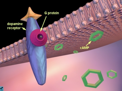
Using the close-up view, explain what happens when dopamine binds to its receptor. When dopamine binds to its receptor, another protein called a G-protein (in pink) moves up close to the dopamine receptor. The G-protein signals an enzyme to produce cyclic adenosine monophosphate (cAMP) molecules (in green) inside the cell. [Sometimes the signal can decrease production of cAMP, depending on the kind of dopamine receptor and G-protein present.] Point to the dopamine receptor-G-protein/adenylate cyclase complex, and show how cAMP is generated when dopamine binds to its receptor. Indicate that cAMP (point to the cyclic-looking structures) controls many important functions in the cell including the ability of the cell to generate electrical impulses.
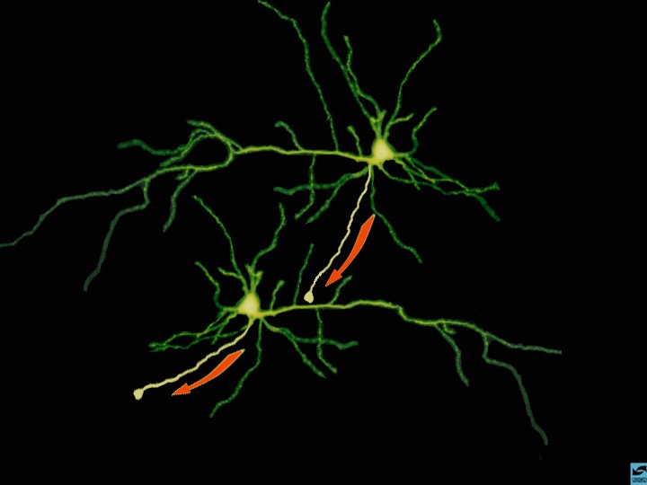
- Summary of neuronal transmission
-
Image

Use the example of two neurons making contact to summarize neuronal transmission. Point to the cell on the top and indicate that electrical impulses flow in the direction toward the terminal. Remind the students what happens when impulses reach the terminal; neurotransmitters are released, they bind to their receptors, and new impulses are generated in the cell on the bottom. Explain that this is how information travels from neuron to neuron.


Introduction to the Reward System
- Reward: drug self-administration
-
Image
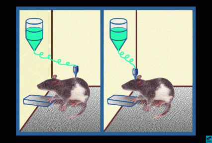
Introduce the concept of positive reinforcement or reward. Explain that rats will press a lever to self-administer an injection of cocaine or heroin that is inserted into either the peripheral bloodstream (left image) or into specific brain regions (right image). The rat keeps pressing to get more cocaine or heroin because the drugs make the rat feel so good. This is called positive reinforcement, or reward. Natural rewards include food, water, and sex - each is required to maintain survival of our species. Animals and people will continue to exhibit a behavior that is rewarding, and they will cease that behavior when the reward is no longer present. Explain that there is actually a part of the brain that is activated by natural rewards and by artificial rewards such as addictive drugs. This part of the brain is called the reward system. Neuroscientists have been able to pinpoint the exact parts of the brain involved, with the help of the rats. Point to the cartoon on the right and explain that rats will also self-administer addictive drugs directly into their brains, but only into a specific area of the reward system. If the injection needle is moved less than a millimeter away from this crucial area, the rat won't press the lever for more drug. So based on information from working with the rats, scientists have drawn a map of the brain, and located the structures and pathways that are activated when an addictive drug is taken voluntarily. Tell the students that you will show them this "map."
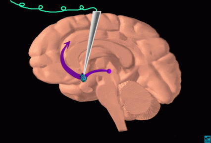
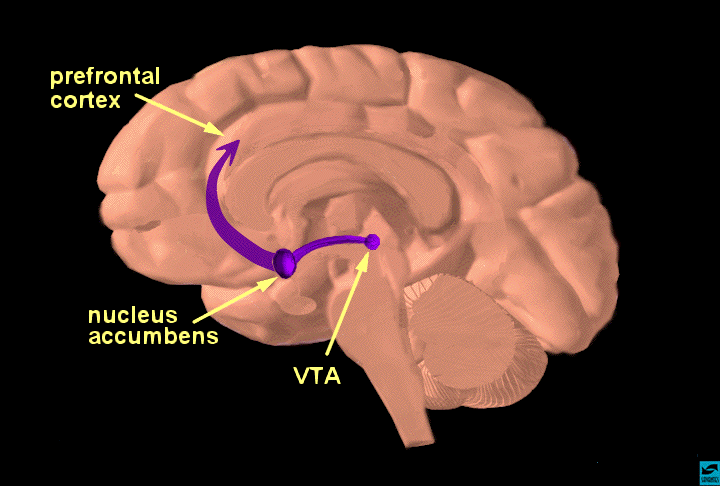
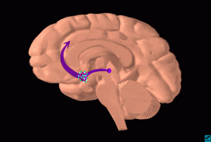
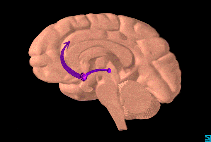
- The reward pathway
-
Image

Tell students that this is a view of the brain cut down the middle. An important part of the reward system is shown and the major structures are highlighted: the ventral tegmental area (VTA), the nucleus accumbens (nuc. acc.) and the prefrontal cortex. Also, the pathway connecting these structures is highlighted. The information travels from the VTA to the nucleus accumbens and then up to the prefrontal cortex. Reiterate that this pathway is activated by a rewarding stimulus. [Note to scientists - this is not the only pathway activated by reward, other structures are involved too, but only this part of the pathway is shown for simplicity.]
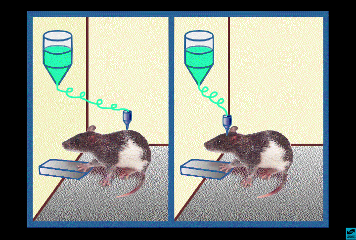
- Injection of cocaine into the nucleus accumbens
-
Image
Demonstrate how scientists located the structures important for the addictive nature of drugs. Show that a rat will self-administer cocaine directly into the nucleus accumbens (or the VTA) to activate the pathway. Point to an area close to the nucleus accumbens or VTA and state that if the injection is placed in this other area, the rat will not press the lever to receive the drug. Indicate that scientists know a lot more than where the drug acts to produce rewarding effects - they also know how the drugs work. Show examples with cocaine, heroin, and marijuana.

Introduction to Drugs of Abuse: Cocaine, Opiates (Heroin) and Marijuana (THC)
- Localization of cocaine binding sites
-
Image
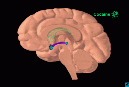
When a person smokes or snorts cocaine, it travels quickly to the brain. Although it reaches all areas of the brain, it concentrates in some specific areas. These are highlighted with the turquoise sprinkles; the VTA, the nucleus accumbens, and the caudate nucleus (lighter turquoise since the caudate is inside the hemisphere). Point out that cocaine concentrates especially in the reward areas that you have just discussed. Cocaine accumulation in other areas such as the caudate nucleus can explain other effects such as increased stereotypic behaviors (pacing, nail-biting, scratching, etc..)
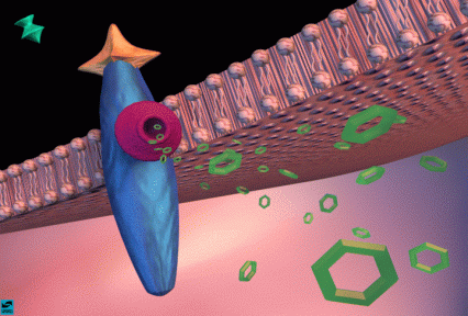
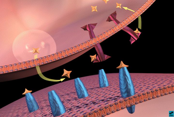
- Dopamine binding to receptors and uptake pumps in the nucleus accumbens
-
Image

Explain that cocaine concentrates in areas of the brain that are rich in dopamine synapses. Review dopamine transmission in the nucleus accumbens. Point to dopamine in the synapse and to dopamine bound to dopamine receptors and to uptake pumps on the terminal.
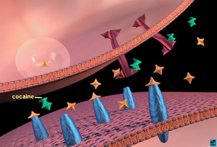
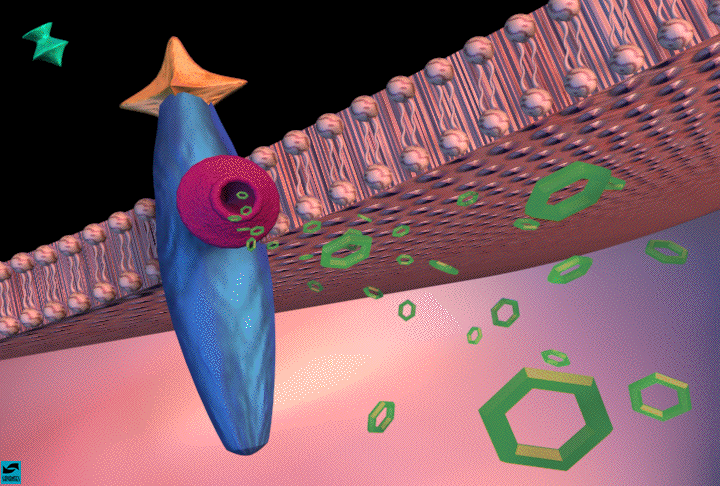
- Cocaine binding to uptake pumps: inhibition of dopamine uptake
-
Image
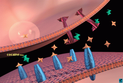
Now, show what happens when cocaine is present in the synapse. Cocaine (turquoise) binds to the uptake pumps and prevents them from removing dopamine from the synapse. This results in more dopamine in the synapse, and more dopamine receptors are activated.
- Increased cAMP produced in post-synaptic cell
-
Image
In a closer view, show how this affects the function of the cell. The increased activation of dopamine receptors causes increased production of cAMP inside the post-synaptic cell. This causes many changes inside the cell that lead to abnormal firing patterns.
- Summary - cocaine binding in nucleus accumbens and activation of reward pathway
-
Image

Show the "big picture," As a result of cocaine's actions in the nucleus accumbens (point to the sprinkles of cocaine in the nuc. acc.), there are increased impulses leaving the nucleus accumbens to activate the reward system. Indicate that with continued use of cocaine, the body relies on this drug to maintain rewarding feelings. The person is no longer able to feel the positive reinforcement or pleasurable feelings of natural rewards (food, water, sex).
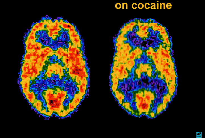
- Positron emission tomography (PET) scan of a person on cocaine
-
Image
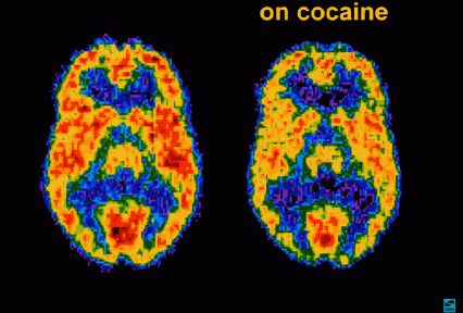
Cocaine has other actions in the brain in addition to activating reward. Scientists have the ability to see how cocaine actually affects brain function in people. The PET scan allows one to see how the brain uses glucose; glucose provides energy to each neuron so it can perform work. The scans show where the cocaine interferes with the brain's use of glucose - or its metabolic activity. The left scan is taken from a normal, awake person. The red color shows the highest level of glucose utilization (yellow represents less utilization and blue shows the least). The right scan is taken from a cocaine abuser on cocaine. It shows that the brain cannot use glucose nearly as effectively - show the loss of red compared to the left scan. There are many areas of the brain that have reduced metabolic activity. The continued reduction in the neurons' ability to use glucose (energy) results in disruption of many brain functions.
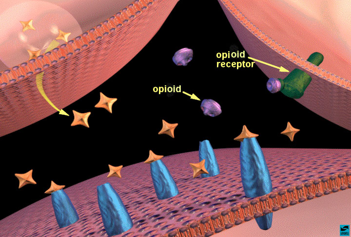
- Localization of opioid binding sites
-
Image
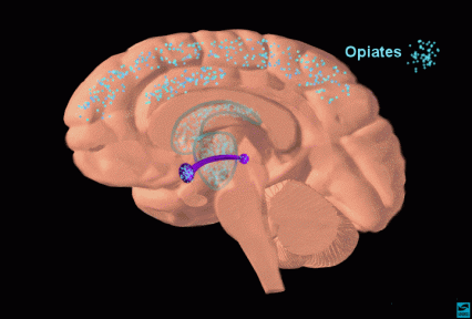
When a person injects heroin or morphine, it too, travels quickly to the brain. Point to the areas where opioids concentrate. The VTA, nucleus accumbens, caudate nucleus and thalamus are highlighted. The opioids bind to opioid receptors that are concentrated in areas within the reward system. Indicate that the action of opioids in the thalamus contributes to their ability to produce analgesia.
- Opioids binding to opioid receptors in the nucleus accumbens: increased dopamine release
-
Image

Show how opioids activate the reward system using the nucleus accumbens as an example. Explain that the action is a little more complicated than cocaine's because more than two neurons are involved. Point out that 3 neurons participate in opioid action; the dopamine terminal, another terminal (on the right) containing a different neurotransmitter (probably GABA for those that would like to know), and the post-synaptic cell containing dopamine receptors. Show that opioids bind to opioid receptors (green) on the neighboring terminal and this sends a signal to the dopamine terminal to release more dopamine. [In case an inquisitive student asks how - one theory is that opioid receptor activation decreases GABA release, which normally inhibits dopamine release - so dopamine release is increased.]
- Increased cAMP produced in post-synaptic cell
-
Image
In a closer view, again, show how this affects the function of the post-synaptic cell. Since there is more dopamine released, there is increased activation of dopamine receptors, similar to the effect of cocaine. This causes increased production of cAMP inside the post-synaptic cell, which alters the normal activity of the neuron.
- Summary - opioid binding in nucleus accumbens and activation of the reward pathway
-
Image

Show the "big picture". As a result of opioid actions in the nucleus accumbens (point to the sprinkles of opioids in the nuc. acc.), there are increased impulses leaving the nucleus accumbens to activate the reward system (point to the frontal cortex). As with cocaine, continued use of opioids makes the body rely on the presence of the drug to maintain rewarding feelings and other normal behaviors. The person is no longer able to feel the benefits of natural rewards (food, water, sex) and can't function normally without the drug present.
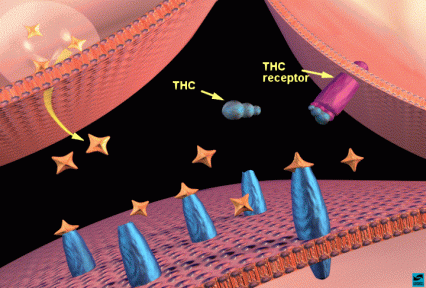
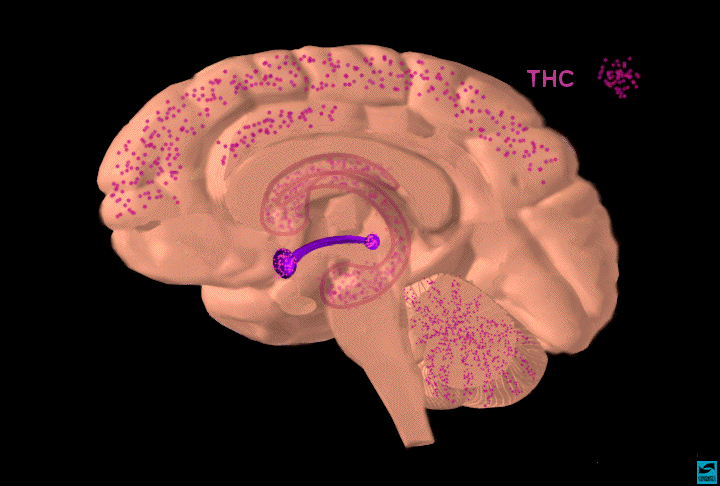
- Localization of THC binding sites
-
Image

When a person smokes marijuana, the active ingredient, THC, travels quickly to the brain. Point to the areas where THC (magenta) concentrates. The VTA, nucleus accumbens, caudate nucleus, hippocampus, and cerebellum are highlighted. THC binds to cannabinoid receptors that are concentrated in areas within the reward system as well as these other areas. Indicate that the action of THC in the hippocampus explains its ability to interfere with memory and actions in the cerebellum are responsible for its ability to cause incoordination and loss of balance. Just as the body produces and responds to its own opioids (endorphins), the body produces and responds to its own cannabinoid molecules, called endocannabinoids. THC interacts with the body’s endocannabinoid signaling system.
- THC binding to THC receptors in the nucleus accumbens: increased dopamine release
-
Image
Over the last few decades, there has been intense study to learn about the endocannabinoid system, not only because of the popularity of marijuana as a recreational drug but also because of the increased use of marijuana for medical purposes. THC is thought to act in a similar way to opioids. Again, use the nucleus accumbens as an example. The same 3 neurons are probably involved; the dopamine terminal, another terminal (on the right) containing a different neurotransmitter (GABA), and the post-synaptic cell containing dopamine receptors. Ask the students if they can tell you how THC might work. THC binds to THC receptors (magenta) on the neighboring terminal and this inhibits the GABA signal, which causes the dopamine terminal to release more dopamine.
- Increased cAMP produced in post-synaptic cell
-
Image
In a closer view, show how this affects the function of the post-syanaptic cell. Since there is more dopamine released, there is increased activation of dopamine receptors. This causes increased production of cAMP inside the post-synaptic cell which alters the normal activity of the neuron.
- Summary - THC binding in nucleus accumbens and activation of the reward pathway
-
Image

Show the "big picture." As a result of THC actions in the nucleus accumbens (point to the concentration of THC in the nuc. acc.), there are increased impulses leaving the nucleus accumbens to activate the reward system (point to the frontal cortex). Scientists still don't know how the continued use of marijuana alters the reward system. Indicate that this is an area of intense research by neuroscientists.
- Overall summary - these drugs of abuse all activate the reward system via increasing dopamine neurotransmission
-
Image
In this last slide, the binding of all 3 drugs is shown in one of the reward areas, the nucleus accumbens. Summarize that each drug increases the activity of the reward pathway by increasing dopamine transmission. This happens even though the drugs act by different mechanisms. Because of the way our brains are designed, and because these drugs activate a particular brain pathway for reward, they have the ability to be misused.
Start a discussion; ask the students if they can think of any other drugs that are misused that probably activate the reward system in the same way. Answer: alcohol, nicotine, and amphetamine are good examples.


Additional Guidance
- Background Information for the Presenter
-
Objectives
The objective of the teaching packet is to inform students (high school) how 3 drugs of abuse (cocaine, opiates, marijuana) actually work in the brain. The packet is arranged in 3 sections. The first section introduces the brain and presents some basic neurobiology, the second introduces the reward pathway and the third presents the mechanism of action of each drug and how each affects the reward system.
Before Entering the Classroom
- Arrange to talk with the teacher (by telephone or in person).
- Obtain information on the age group and extent of biology background.
- Discuss the subjects that you will present to the class.
- If possible, meet with the teacher & review the presentation; provide teacher with some background material if requested.
- Solicit advice from the teacher about teaching strategies (e.g. what to do if some students are disruptive or not listening).
- Read the narrative script and practice the presentation - this is different from a scientific seminar to peers!
Once in the Classroom
- Introduce yourself to the class.
- Talk briefly about your path to your current job.
- Describe what you do (simply).
- Indicate the object of your presentation.
- Invite questions during your presentation.
- If the opportunity arises, ask questions of the students.
- Be prepared to define any words you use.
General Instructions
- The presentation should take approximately 30-40 minutes (without questions); it can be presented in 2 class sessions if so desired.
- Use the narrative text as a guide, it need not be repeated word-for-word.